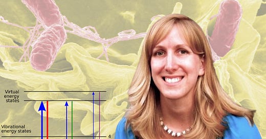
For a patient with sepsis (an infection in the blood), the chance of survival decreases by 7-10% every hour. It’s important to quickly identify what bacteria are present and which antibiotics might be effective.
Typically, there are very few bacteria in the blood. Culturing bacteria from a sample can take 12-24 hours and then one needs to test for drug susceptibility. Speeding up this process would save a lot of lives.
Jen Dionne and her collaborators at Stanford are working on this problem using Raman spectroscopy. Through a combination of nanophotonics, acoustic bioprinting, and machine learning, they are developing methods to quickly and sensitively identify bacteria and screen for antibiotic susceptibility.
What is Raman Spectroscopy?
Raman spectroscopy is basically inelastic photon scattering. So if you think about having a laser or some other monochromatic light source, you shine that onto your sample, say onto a cell, like a bacterial cell. And the molecular vibrations in that sample basically add to or subtract from the energy of the laser source.
You wind up with a fingerprint or a series of scattered wavelengths that are different from your incident wavelength. And you can use that spectrum to identify the constituents of what's in your sample.
Raman spectra of complex mixtures appear as a wavy line, more like a snapshot of an oscillating jump rope than a series of distinct peaks that you would get from a pure sample of a small molecule.
Here is what you need to know:
It’s possible to do Raman spectroscopy on whole cells. By training machine learning models on thousands of bacterial samples, Raman can identify different bacterial species and determine which ones are resistant to specific antibiotics.
Raman is typically not very sensitive but the efficiency goes up by the 4th power of the electric field strength. Surface Enhanced Raman Spectroscopy (SERS) using nanoparticles made of superconducting materials can focus light energy to a very small region without heating and destroying the sample, boosting the sensitivity. The added sensitivity allows the collection of more data (better resolution, more specificity).
Spectra can be collected in flight as droplets (of blood for example) are acoustically ejected at kilohertz frequencies and printed on a substrate. The identification of bacteria by the spectra can be confirmed by electron microscopy of printed drops on the substrate.
Rapid diagnosis of sepsis is just one possible application. Analysis of environmental samples and wastewater epidemiology are also possible with this method.
The takeaway for me is that with a large enough data set of samples, machine learning algorithms can identify subtle features that would never be possible for a human being. Adding more data through nanophotonics opens up even more possibilities to tackle bigger problems.
Your deepest insights are your best branding. I’d love to help you share them. Chat with me about custom content for your life science brand.
Cover Image Credit (energy states): Moxfyre, based on work of User:Pavlina2.0, CC BY-SA 3.0, via Wikimedia Commons


Share this post